The Evolution of Emergency Gastrointestinal Procedures: How Robotic Surgery Enhances Outcomes in Acute Appendicitis

Emergency gastrointestinal surgery has undergone a profound transformation over the past several decades. Historically, acute surgical emergencies of the abdomen required large open incisions, significant tissue disruption, and prolonged hospital stays. Among these emergencies, acute appendicitis remains the most common cause of sudden, severe abdominal pain requiring immediate surgical intervention.
As medical technology has advanced, the surgical management of appendicitis has transitioned from conventional open appendectomy to laparoscopic techniques, and now to computer-assisted, robot-assisted surgical systems. Understanding the clinical progression from open surgery to modern robotic surgery demonstrates how surgical innovation continues to improve patient safety, clinical precision, and recovery trajectories in acute appendicitis management.
The Clinical Pathology of Acute Appendicitis
Acute appendicitis is an acute inflammatory condition affecting the vermiform appendix, a vestigial tubular structure located at the confluence of the cecum in the right lower quadrant of the abdomen. The pathogenesis of acute appendicitis typically begins with obstruction of the appendiceal lumen. Luminal blockage can result from a variety of mechanical and biological factors, including:
- Fecaliths: Hardened, calcified deposits of fecal matter that occlude the narrow opening of the appendix.
- Lymphoid Hyperplasia: Reactive swelling of the submucosal lymphoid tissue, frequently associated with localized or systemic viral or bacterial infections.
- Foreign Bodies or Neoplasms: Less frequent causes of mechanical occlusion that impede fluid drainage.
Following luminal blockage, normal mucosal secretions accumulate distal to the obstruction, causing a rapid increase in intraluminal pressure. This pressure compromises venous and lymphatic circulation within the appendiceal wall, leading to mucosal ischemia, bacterial transmural invasion, and tissue necrosis. If the inflammatory cascade is not interrupted by prompt surgical excision, the ischemic appendiceal wall will rupture, releasing purulent and fecal material into the peritoneal cavity. Perforation can lead to localized intra-abdominal abscesses, extensive phlegmon formation, or generalized peritonitis-a life-threatening systemic infection.
The Historical Paradigm Shift in Surgical Management
To appreciate the impact of robotic surgery on modern emergency care, it is useful to examine the three distinct eras of appendectomy techniques.
1. The Open Appendectomy Era
For nearly a century following its introduction in the late 19th century, open appendectomy was the gold standard for treating acute appendicitis. The procedure involved making an incision in the right lower quadrant-commonly a McBurney or Davis-Rockey incision-dividing abdominal muscle fibers, directly visualizing the cecum, ligating the appendiceal artery, and resecting the inflamed organ. While effective at removing the source of infection, open surgery carries inherent drawbacks:
- Higher postoperative pain levels due to muscle-splitting abdominal wall incisions.
- Increased incidence of surgical site infections, particularly in complex or perforated cases.
- Longer recovery times before returning to physical activity and work.
- Greater risk of post-surgical incisional hernias and peritoneal adhesions.
2. The Laparoscopic Revolution
Introduced in the late 20th century, laparoscopic appendectomy fundamentally altered gastrointestinal surgery. By utilizing a camera (laparoscope) and long, rigid instruments inserted through three small trocars (ranging from 5 to 12 millimeters), surgeons could visualize and excise the appendix without large abdominal incisions. Laparoscopic surgery demonstrated clear clinical benefits, including reduced wound complication rates, decreased postoperative pain, shorter hospital stays, and quicker functional recovery.
However, standard laparoscopy possesses inherent technological limitations:
- Fixed Instruments: Rigid, non-articulating instruments limit range of motion inside tight anatomical spaces.
- Two-Dimensional Vision: Standard laparoscopic displays provide 2D visualization, requiring surgeons to infer depth perception.
- Fulcrum Effect: Operating through fixed abdominal wall ports creates counter-intuitive hand movements and increases surgeon fatigue.
How Robotic Surgery Refines Emergency Gastrointestinal Procedures
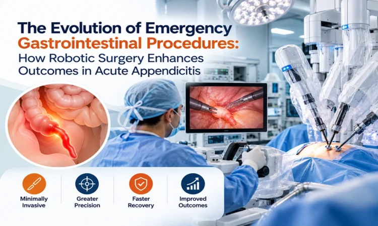
Robotic-assisted surgery represents the latest evolution in minimally invasive abdominal intervention. While the term “robotic” implies automated execution, the system is strictly a surgeon-controlled tele-manipulative platform. The surgeon sits at an ergonomic console adjacent to the operating table, translating hand movements into precise, micro-instrument manipulations inside the patient’s body.
When applied to the management of acute appendicitis-especially complex, gangrenous, or retrocecal presentations-robotic surgery offers significant technological and clinical enhancements over standard approaches.
1. High-Definition 3D Visualization and Magnification
Robotic platforms provide high-definition, three-dimensional spatial visualization with up to 10x magnification. Unlike the flat 2D view of standard laparoscopy, 3D visualization allows the surgical team to clearly differentiate delicate tissue planes, identify appendiceal vascular structures, and evaluate neighboring pelvic organs. This visual clarity is particularly valuable when the appendix is inflamed, retrocecal (positioned behind the cecum), or bound by dense inflammatory adhesions.
2. Wristed Instrumentation and Superior Dexterity
Standard laparoscopic tools are straight and offer only four degrees of freedom. In contrast, robotic instruments feature articulated wrists (EndoWrist technology) that emulate the human hand and wrist, providing seven degrees of freedom and 90 degrees of articulation. This enhanced dexterity allows surgeons to perform complex dissection, tissue retraction, and intracorporeal knot tying within confined pelvic geometry that would otherwise require open surgical conversion.
3. Motion Scaling and Tremor Filtration
Robotic control systems actively filter out physiological hand tremors and allow motion scaling. For instance, a three-centimeter movement of the surgeon’s hand at the control console can be scaled down to a precise one-centimeter movement of the instrument tip inside the abdominal cavity. This steady precision minimizes accidental collateral tissue trauma, reduces micro-vascular bleeding, and protects adjacent structures such as the ureter and iliac vessels.
4. Advanced Real-Time Tissue Assessment
Modern robotic platforms incorporate advanced intraoperative imaging technologies, such as near-infrared fluorescence imaging using indocyanine green dye. This capability allows real-time visualization of tissue perfusion and vascular anatomy. In severe acute appendicitis, assessing the viability of the appendiceal base and adjacent cecal tissue helps surgeons decide whether simple ligation is sufficient or if cecal closure techniques are needed to prevent postoperative stump leaks.
Clinical Outcomes and Patient Benefits
The technological superiority of robotic surgery translates directly into improved clinical metrics and patient outcomes in acute appendicitis cases:
- Reduced Conversion Rates to Open Surgery: In difficult appendicitis cases marked by severe inflammation, retrocecal positioning, or anatomical variation, robotic dexterity allows surgeons to complete the resection minimally invasively, avoiding large open incisions.
- Decreased Risk of Complications: Enhanced visual accuracy and gentle tissue handling reduce intraoperative hemorrhage, accidental bowel deserosalization, and post-operative intra-abdominal abscess formation.
- Minimization of Postoperative Discomfort: By reducing stress and torque on the abdominal wall ports, robotic platforms cause less abdominal wall trauma. Patients routinely report lower visual analog scale pain scores and require significantly fewer opioid analgesics postoperatively.
- Accelerated Functional Recovery: Smaller incisions and reduced systemic surgical stress lead to faster return of bowel function (peristalsis), earlier oral fluid intake, and shorter hospital stays. Most patients undergoing uncomplicated robotic appendectomy are discharged within 24 hours and resume normal daily routines within a week.
Conclusion
The evolution of emergency gastrointestinal surgery reflects a continuous push toward minimizing surgical trauma while optimizing diagnostic and therapeutic precision. From the large open incisions of the past to the refined access of laparoscopy, each technical breakthrough has advanced patient safety. Today, robotic surgery represents a major milestone in managing acute appendicitis. By combining 3D high-definition visualization, wristed multi-directional instrumentation, and steady digital control, robotic systems enable surgeons to navigate complex acute abdominal pathology with high precision, yielding fewer surgical complications, lower pain levels, and faster recovery times for patients.